
High-resolution 3D microscopy is performed routinely at synchrotron radiation facilities in Europe, with applications in a range of bio-medical research proplems. In this workshop, expert synchrotron users and staff will present state-of-the-art case studies and we will discuss future possibilities both at MAX IV and abroad.
Participants will be welcomed to the workshop by Lund University Vice-Chancellor Erik Renström.
See the lineup of speakers below – more to be confirmed!
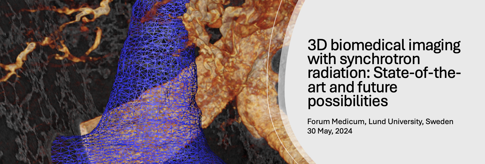
Title: Workshop: 3D biomedical imaging with synchrotron radiation: State-of-the-art and future possibilities
When: 30 May, 2024
Venue: Utblicken (room E 16), top floor Forum Medicum, Sölvegatan, Lund University, Sweden, or online via Zoom. A Zoom-link will be sent to all digital participants in time for the event.
Registration info: Please note that there is a limited number of available spaces for physical participants, and the registration for in-person participation will close when the venue is full! We also advise all participants to book accommodation as soon as possible, since this is generally a busy week in Lund.
Confirmed Speakers

Other info
Participation fee:
600 SEK for physical participation incl 3-course dinner at Kulturen's restaurant
400 SEK for physical participation without dinner
200 SEK for online participation (via Zoom)
-
The program may still be subject to some change
30 May
Morning (circa 10–11, exact time will be announced soon): Optional tour to MAX IV for registered participants
11:30–12:50 Lunch and registration, Venue: E16 “Utblicken”, top floor of Forum Medicum, Sölvegatan, Lund
12:50–13:00 Welcome by Vice-Chancellor Erik Renström
13:00–13:10 Welcome and introduction, Martin Bech (LINXS/HALRIC)
Session 1 Chair: Marco Stampanoni, Paul Scherrer Institut, Switzerland
13:10–13:25 Title TBA, Alexandra Pacureanu, ESRF
13:25–13:40 Title TBA, Anne Bonnin, Paul Scherrer Institut, Switzerland
13:40–13:55 "3D imaging of pulmonary vascular disease" Karin Tran-Lundmark, Lund University, Sweden
13:55–14:10 Title TBA, Goran Lovric Paul Scherrer Institut, Switzerland
14:10–14:25 Leg stretcher
Session 2 Chair: Kristina Djinovic Carugo, EMBL, France
14:25–14:40 “Hard X-ray nano-imaging of Toxoplasma gondii in different development stages”, Matthew Bowler, EMBL, France
14:40–14:55 Title TBA, Sam Bayat, Grenobles Alpes University, France
14:55–15:10 “3D virtual histology with synchrotron and advanced laboratory radiation: a new tool for clinical research and pathology”, Tim Salditt, Universität Göttingen, Germany
15:10–15:25 Title TBA, Maximilian Ackermann, Johannes Gutenberg University Mainz, Germany
15:25–15:30 Exciscope flash presentation
15:30–16:00 Coffee break
Session 3 Chair: Marjolein Thunnisen, MAX IV Laboratory
16:00–16:15 Title TBA, Henrik Birkedal, Aarhus University, Denmark
16:15–16:30 “Investigation of cardiomyopathy using X-PCI (deep geno-phenotype of heart muscle disease)” Kan Yan Chloe Li, University College London, UK
16:30–16:45 Title TBA, Isabel Gonçalves, Lund University, Sweden
16:45–17:30 Panel discussion and wrap-up session
19:00– Optional dinner at Restaurant Kulturen in Lund
-
Martin Bech (Lund University), Kristina Djinovic (EMBL Grenoble), Trevor Forsyth (LINXS, Sweden), Michael Krisch (ESRF, France), Marco Stampanoni (PSI, Switzerland), Marjolein Thunnisen (MAX IV, Sweden), Karin Tran Lundmark (Lund University)
-
3D virtual histology with synchrotron and advanced laboratory radiation: a new tool for clinical research and pathology
Tim Salditt,
Institut für Röntgenphysik, Universität Göttingen, and DFG-Excellence Cluster 2067 Multiscale Bioimaging
In order to unravel physiological and pathological mechanisms at the cellular level, structure and processes have to be visualized on a wide range of scales. Imaging at cellular and sub-cellular resolution is the realm of histology. For this purpose, the tissue obtained by surgical intervention or from a post mortem autopsy is cut into thin sections, stained and observed in an optical microscope. In conventional histology, images are obtained only of two-dimensional sections but not of the entire three-dimensional (3D) volume. In order to visualise and to quantify the cytoarchitecture in 3D, even deep in the tissue or organ, we use phase-contrast X-ray computerized tomography , as a tool for quantitative and fully digital 3D virtual histology [1]. We have implemented the method using optimized phase retrieval [2,3], both at highly coherent synchrotron and at inhouse micro-focus sources. In a multi-scale approach, we combine parallel and cone beam illumination to cover a wide range of scales. Since the workflow is non-destructive and fully compatible with standard clinical pathology, we can perform correlative histology studies.
In this talk we discuss image formation and advanced phase retrieval of propagation and inline holography data, the respective resolution limits, object constraints, scaling properties, as well as morphometric image analysis. We show how solutions and algorithms of mathematics of inverse problems and machine learning [2-4] help us to meet the challenges of phase retrieval, tomographic reconstruction, segmentation, and more generally exploitation of bulky image data. All to the benefit of ambitious imaging projects such as mapping the human brain [4,6] of fighting infectious diseases [6].
References:
[1] T. Salditt, A. Egner and R. D. Luke (Eds.)
Nanoscale Photonic Imaging
Springer Nature (2020), TAP, 134, Open Access Book
[2] L. M. Lohse, A.-L. Robisch, M. Töpperwien, S. Maretzke, M. Krenkel, J. Hagemann and T. Salditt
A phase-retrieval toolbox for X-ray holography and tomography
Journal of Synchrotron Radiation (2020), 27, 3
[3] S. Huhn, L.M. Lohse, J. Lucht, T. Salditt
Fast algorithms for nonlinear and constrained phase retrieval in near-field X-ray holography based on Tikhonov regularization - arXiv preprint arXiv:2205.01099 (2022)
[4] M. Eckermann, B. Schmitzer, F. van der Meer, J. Franz, O. Hansen, C. Stadelmann and T. Salditt
Three-dimensional virtual histology of the human hippocampus based on phase-contrast computed tomography
Proc. Natl. Acad. Sci. (2021), 118, 48, e2113835118
[5] M. Eckermann, J. Frohn, M. Reichardt, M. Osterhoff, M. Sprung, F. Westermeier, A.Tzankov, C. Werlein, M. Kuehnel, D. Jonigk and T. Salditt
3d Virtual Patho-Histology of Lung Tissue from Covid-19 Patients based on Phase Contrast X-ray Tomography
eLife (2020), 9:e60408
[6] M. Reichardt, P.M. Jensen, V.A. Dahl, A.B. Dahl, M. Ackermann, H. Shah, F. Länger, C. Werlein, M.P. Kuehnel, D. Jonigk and T. Salditt
3D virtual histopathology of cardiac tissue from Covid-19 patients based on phase-contrast X-ray tomography
eLife (2021), 10:e71359
Contact:
For practical questions about the workshop, please contact josefin.martell@linxs.lu.se
The workshop is organised in the context of the LINXS IPDD theme.
Sponsors

During our events we sometimes take photographs and short film clips to profile our activities. Please let us know if you don’t want to be in any photos/films before we start the event. Some webinars are recorded to be used for educational purposes in the LINXS website.
By registering to our events you give your permission to LINXS, according to the General Data Protection Regulation (GDPR), to register your name and e-mail address to be used for the sole purpose of distributing newsletters and communications on LINXS activities.
If you don’t receive registration confirmation please check your spam mail.