
In search of nutritious, sustainable and more resilient oats - research made possible by the NLF theme
This is a story about oats – and research to make oats more nutritious, resilient and versatile. It is also a story about what lies at the heart of all scientific discoveries – passion, curiosity, hard work and collaboration, all made possible by the Northern Lights on Food theme at LINXS, with funding from Tillväxtverket - Swedish Agency for Economic and Regional Growth.
The health benefits of oats have been known for centuries: the fiber and protein content contribute towards feeling of fullness, as well as slower blood glucose release. Oats are also a source of vitamins and minerals such as magnesium, copper, thiamine and zinc. Yet, as populations are facing increasing challenges related to food scarcity, malnutrition, obesity, and climate change, the need to develop oat seeds that are more nutritious, easier to digest and of higher quality is increasing.
Today, it is simply not enough to only grow regular oats, we need to select oats that are better for both human health and the planet, says Nick Sirijovski, who completed his PhD thesis at Lund University, and now works for Oatly.
In early 2023, with the support by the Northern Lights on Food theme at LINXS, Nick joined forces with his former supervisor, Mats Hansson, Professor at the Department of Biology at Lund University, and tomography expert Emanuel Larsson, also at Lund University. Their aim? To find out as much as possible about the composition of different oat seeds, in particular the 3D distribution of beta glucan, which is the most soluble fibre of oat.
A long journey to create a favorable sample scanning conditions
The group began the process by performing lab-based X-ray microtomography (µCT) experiments at the 4D Imaging Lab at Lund University. The purpose was to ascertain what imaging methods to use to resolve highly detailed images of the internal structure of the oat. At first, the experiments did not yield the hoped-for results. Scanning the entire oat sample with a broader Field of View (FOV) did not provide the resolution needed to resolve the target structures. After cutting the sample, to increase the contrast and resolution, while reducing the FOV, the sample instead dried out in the µCT scanner, which can reach a temperature of 25 degrees. This happened frequently during the longer experiments.

To the left: demonstration of various methods to fixate the seed samples prior to scanning with lab-based X-ray microtomography. A) entire seed fixated inside a plastic tube. B) cut seed fixate on the top of a cut tube and C) with surrounding parafilm. D) cut seed fixated inside a plastic tube and immersed in PTA liquid. To the left: the photo shows a mounted PTA-stained seed inside a plastic tube and immersed in PTA liquid. The sample is about to be scanned inside the X-ray lab tomograph at the 4D Imaging Lab at Lund University. The blue colour represents the path of the incoming X-ray cone beam.
– We spent months trying to create a stable, favorable sample condition for the oat seeds. When we started, we did not think it was going to take this long. It is a bit like detective work: you have to try many different things, says Emanuel Larsson, researcher at the Department of Experimental Medical Science at the Faculty of Medicine, Lund University and Coordinator for CIPA – Correlative Image Processing and Analysis – a cross-faculty infrastructure at Lund University.
Staining the oat seed with metal ions yielded results
After some time, Emanuel, Nick and Mats came up with a solution: to put parafilm around the sample to make it withstand the heat inside the scanner. But they soon realised that the problem was not solved. The images did not show enough contrast needed to be able to segment and analyse the image data. After long discussions back and forth, they came up with another approach: to stain the oat seed with phosphotungstic acid (PTA).
– I had read about this technique being used in biology, so we decided to try it. We stained the sample by letting them soak in PTA for a number of weeks, then we cleaned them with buffer solution, fixated them inside a plastic tube and added fresh PTA solution, and sealed with parafilm on the top. By scanning the seeds surrounded by PTA liquid, there was no risk of drying out the samples, says Emanuel Larsson.

2D microscopy images of ot seeds. A) light microscopy of the cell structure. B) Fluorescence microscopy of oat seed, where Beta-glucan has been labelled with a fluorophore in blue.
Finally, the researchers were beginning to see the distribution of the germ, the endosperm and bran – the tree components which make up the oat seed. The starch and the beta glucan are located in the endosperm, the beta glucan is in the outer layers.

Prof. Mats Hansson inspects cut planes and a 3D-rendering of a PTA-stained Oat seed, scanned with X-ray microtomography.
Once we had the first results, I was so excited! They appeared after nine hours in the µCT scanner, and I called Nick straight away! In the images, we could clearly see the endosperm cell wall structures that contain the beta glucan, says Emanuel Larsson.
He reflects that it is the back-and forth dialogue between him and other researchers and industry that makes him excited. The sense of achievement from collaborating on ways to improve the sample environment, trying different scan times, and finally staining the sample is important – and what motivates him in his daily work.
Once the group had managed to produce workable data, they had to overcome the next hurdle: segmenting the data and creating 3D renderings that would show the oat structure in a higher accuracy.
– Since we stained the oats, we actually change the grey-level scale, so classical image segmentation methods, e.g., thresholding might not work anymore, says Emanuel Larsson.

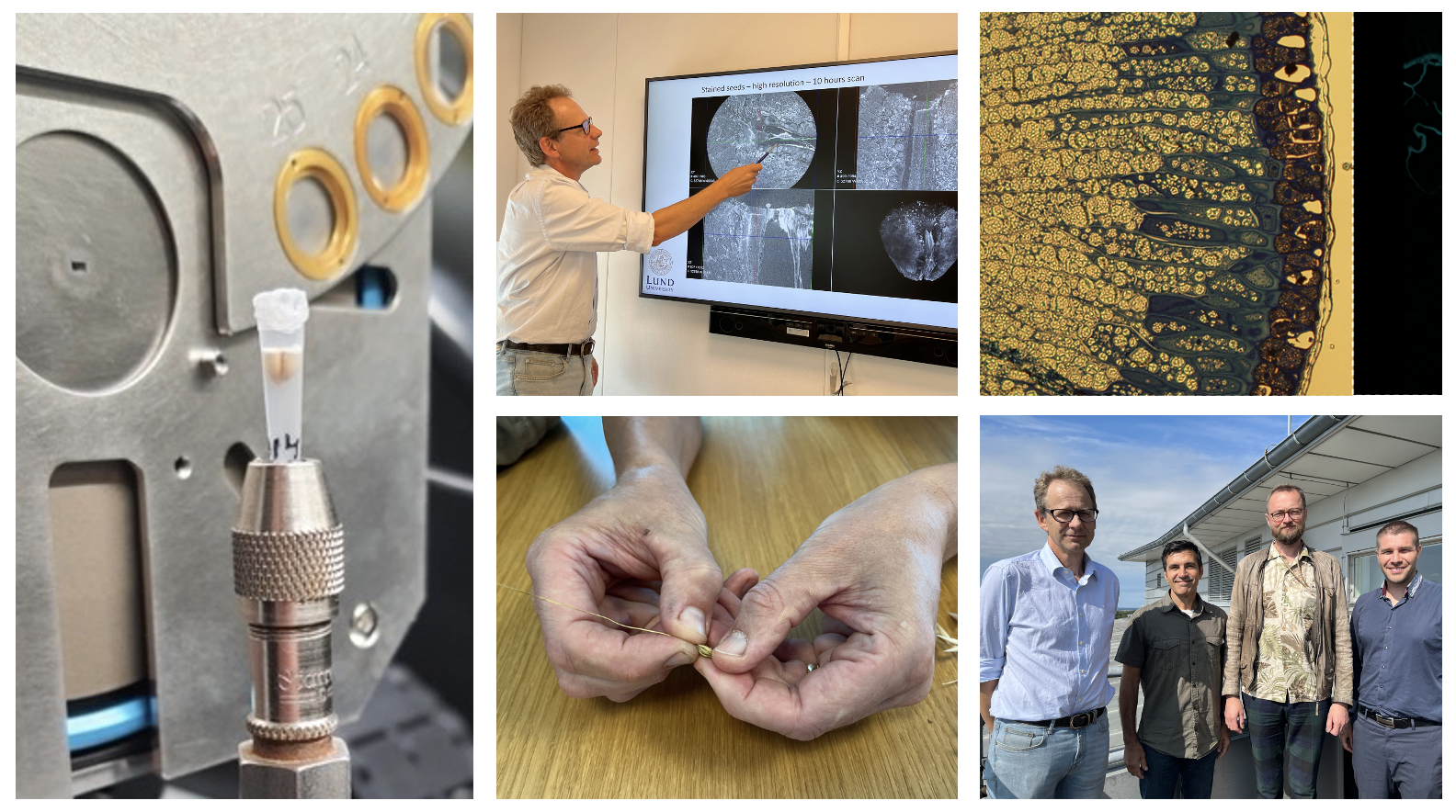
The research team: Professor Mats Hansson, senior lecturer Alexandros Sopasakis, Nick Sirijovski, Oatly, and researcher Emanuel Larsson.
The use of AI made the data analysis better
They made contact with the mathematician Alexandros Sopasakis, senior lecturer at the Centre for Mathematical Sciences at Lund University and application expert at CIPA, who came to their rescue. Alexandros suggested developing a unique AI-model that would take in annotated images, which would be used for training the AI-model to then automatically segment the remaining N number of datasets, which can then be used for 3D rendering and quantitative image analysis. First, the images had to be manually annotated, which was done by having two students manually annotate around 900 images, where they circled the areas of importance, during the summer months. Then they had to teach the system, which Alexandros developed, how to recognize the relevant components within the oat.
The AI-model has now produced its first results: a nicely segmented 3D-model of the oat seed. Alexandros Sopasakis is very happy with the results.
– This work is fascinating for me as a mathematician. The feedback to improve the machine learning, where you go back and forth between the model and the experts is always interesting. This is where the breakthrough happens. Since I had never worked on oats before, I am glad to see that our approach worked.
He continues:
– The worst thing you can do is cold annotating. You need human eyes to tell the machine what it interesting, by for example tracing on the computer screen to show the machine what to recognize. Having students do this job during the summer is a win-win since they get acquainted with the science, while also earning some money, during perhaps their first summer job ever.
Next step is to create a 3D model of different oat grains
Nick, Mats and Emanuel are delighted that they have now completed the first stage of their endeavor to progress basic science on oats. The long-term goal is to perform faster and more highly resolved experiments at a large-scale facility such as MAX IV, which has a dedicated tomography beamline, ForMAX, to create 3D models of different oat grains.
There are so many reasons why this is important. Just think of fractionation processes, if we know more about the 3D distribution of beta glucan, then industry can use that information to improve how they process the seed, says Nick Sirijovski.
He continues:
– Another huge area is plant breeding. The oat genome has recently been sequenced and annotated, which means that there are more possibilities to select completely new oat varieties with optimal seed structure in mind. With this type of research, we can in time connect our data to oat DNA to understand the mechanisms controlling not only variation in total beta glucan content but also its distribution in the seed.
Nick, Emanuel and Mats would also like to see experiments on oats grown in different locations, or exposed to different biotic or abiotic stresses might impact the internal structure of the oat seed.
– You have to start somewhere. And I feel that our collaboration can be the beginning of something solid. If nothing else, our results shows that there is still so much to find out, even though we have grown oats for hundreds of years, says Nick Sirijovski.
The collaboration was supported by Tillväxtverket, EU regionala fonder and Region Skåne.