
VIDEO - CoWork series - Holographic tomography as a new tool for bioimaging and 3d Virtual Patho‐Histology of Lung Tissue from Covid‐19 Patients, with Tim Salditt & Marina Eckermann

VIDEO - CoWork series - Holographic tomography as a new tool for bioimaging and 3d Virtual Patho‐Histology of Lung Tissue from Covid‐19 Patients, with Tim Salditt & Marina Eckermann
The talk explains how the central challenge of inverting the coherent diffraction pattern can be mastered by different reconstruction algorithms in the optical far and nearfield. In particular, it presents the full field projection imaging at high magnification, recorded by illumination with advanced x‐ray waveguide optics and show how imaging and diffraction can be combined to investigate biomolecular structures within biological cells, also correlatively with super‐resolution light microscopy. The talk presents different examples of biophysical and biomedical applications, including 3d virtual histology of human brain tissue..
Speaker: Professor Tim Salditt, the Institute for X-ray Physics of the University of Göttingen and Marina Eckermann is a PhD student in the group of Tim Salditt at the Institute for Xray Physics of the University of Göttingen.
The webinar is part of the LINXS webinar series, CoWork. The CoWork webinar series is dedicated to the exploitation of the coherence properties of X-rays for advanced materials characterization, with a special focus on inverse microscopy techniques, such as Coherent Diffraction Imaging (CDI), Ptychography and Holography.
Abstract
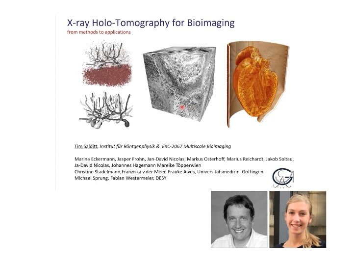
X‐rays can deeply penetrate matter and thus provide information about the functional (interior) architecture of complex samples, from biological tissues and cells to nanoscale composite materials. Until recently, however, this potential of hard x‐rays in view of penetration, spatial resolution, contrast, and compatibility with environmental conditions was significantly limited by the lack in suitable in x‐ray optics. With the advent of highly brilliant radiation, and the development of lens‐less diffractive imaging and coherent focusing, the situation has changed. We now have nano‐focused coherent x‐ray synchrotron beams at hand to probe nanoscale structures both in scanning and in full field imaging and tomography. We explain how the central challenge of inverting the coherent diffraction pattern can be mastered by different reconstruction algorithms in the optical far and nearfield. In particular, we present full field projection imaging at high magnification, recorded by illumination with advanced x‐ray waveguide optics [1,2], and show how imaging and diffraction can be combined to investigate biomolecular structures within biological cells [3], also correlatively with super‐resolution light microscopy. We present different examples of biophysical and biomedical applications [4,5], including 3d virtual histology of human brain tissue [5].
[1] M. Bartels, M. Krenkel, J. Habe, R.N. Wilke, T. Salditt, X‐Ray Holographic Imaging of Hydrated Biological Cells in Solution, Phys. Rev. Lett. 114, 048103 (2015).
[2] L. M. Lohse, A.‐L. Robisch, M. Töpperwien, S. Maretzke, M. Krenkel, J. Hagemann and T. Salditt A phase‐retrieval toolbox for X‐ray holography and tomography Journal of Synchrotron Radiation (2020), 27, 3
[3] M. Bernhardt, J.‐D. Nicolas, M. Osterhoff, H. Mittelstädt, M. Reuss, B. Harke, A. Wittmeier, M. Sprung, S. Köster, T. Salditt. Correlative microscopy approach for biology using x‐ray holography, x‐ray scanning diffraction, and STED microscopy Nat. Comm. (2018), 9, 3641
[4] M. Reichardt, C. Neuhaus, J‐D. Nicolas, M. Bernhardt, K. Toischer and T. Salditt X‐ray structural analysis of single adult cardiomyocytes: tomographic imaging and micro‐diffraction. Biophysical Journal (2020), 119, 7, 1309‐1323
[5] M. Töpperwien, F. Van der Meer, C. Stadelmann, and T. Salditt. Three‐dimensional virtual histology of human cerebellum by X‐ray phase‐contrast tomography, Proceedings of the National Academy of Sciences (2018), 201801678
Biography
Tim Salditt is professor of experimental physics at the Institute for X-ray Physics of the University of Göttingen. He studied physics in Munich and Grenoble, and received his Ph.D. in 1995 for research in kinetic growth of surfaces and interfaces, which he studied by diffuse X-ray scattering and non-specular reflectivity with Hans Peisl at the University of Munich. He then moved his focus to biophysics, using the newly developed approaches for interface-sensitive scattering to study fluctuations in oriented lipid membranes. In his postdoctoral work with Cyrus Safinya in Santa Barbara, he worked on the structure and interactions of lipid/DNA complexes. Driven by the motivation to study biomolecular assemblies also in the hierarchical and complex functional environment of cells, he developed X-ray waveguide optics for nanoscale holographic imaging and tomography. With his group, he has designed a synchrotron radiation instrument, which they operate at the PETRAIII storage ring, together with DESY.
Using a combination of diffraction with micro- and nano-focused beams as well as holographic imaging and tomography, they can now study biomolecular assemblies and biological matter, from the molecular level to biological cells and tissues. Recently, they also got increasingly involved with translating phase contrast tomography from synchrotron to laboratory instrumentation, more amenable for biomedical research groups. They push the limits in X-ray optics, including focusing, wave-front control, coherence, phase retrieval, reconstruction algorithms, information theory and image processing in order to get a maximum of information from a minimum of photons.
Tim Salditt has been spokesperson of the collaborative research center Nanoscale Photonic Imaging for 12 years, and is a principle investigator of the DFG Center for Excellence Multi-Scale Bioimaging: from molecular machines to networks of excitable cells. He is a member of the Academy of Science. In Göttingen, and of the DESY scientific advisory committee.
Marina Eckermann is a PhD student in the group of Tim Salditt at the Institute for Xray Physics of the University of Göttingen. She studied Physics in Göttingen and Munich, with visits in Edmonton and Grenoble. In her Bachelor thesis with Tim Salditt, she investigated the actin-myosin complex in cardiomyocytes using synchrotron-based small-angle X-ray scattering. After an industrial internship in medical technology, she pursued studying physics, with a specialization in Medical Physics. During a research internship at the University of Alberta, Marina Eckermann worked on diffusion analysis based on single-particle tracking in the group of Christopher Cairo.
For her Master thesis, she turned to propagation-based phase-contrast X-ray tomography of neuronal tissues. With Paola Coan, she was a visiting scientist at the ESRF, and collected further experience in synchrotron experiments, as well as in data processing and reconstruction. She stayed in the field, and joined Tim Salditt’s group for her PhD project on 3d virtual histology of neuronal tissues using propagation-based phase-contrast X-ray tomography. As her main project, she studied hippocampal neurons from a number of different human subjects, with a group affected by Alzheimer’s disease. This project again included data collection and processing, and further involved image segmentation techniques, statistical analysis and mathematical approaches from optimal transport theory, to uncover subtle pathological changes. This again comprised synchrotron and home-built laboratory setups. Marina Eckermann is also interested in advanced sample preparation techniques involving metal staining, in correlative experimental methods, and as of last year in using phasecontrast tomography to fight the Covid-19 pandemic.