
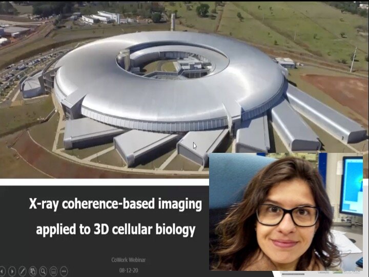
VIDEO - CoWork series - X-ray coherence-based imaging applied to 3D cellular biology, with Carla Cristina Pólo

VIDEO - CoWork series - X-ray coherence-based imaging applied to 3D cellular biology, with Carla Cristina Pólo
X-ray coherence-based imaging comprises several powerful techniques which allow investigations in the different scientific fields, including biology and life sciences. The non-crystalline nature of such samples can be explored by coherent diffractive imaging (CDI) and ptychography and cellular biology has benefited from such techniques to unveil cellular architecture of bacteria, yeast and parasites at high resolutions and in 3D. However, studies with tissues involves technical challenges during sample preparation and data collection, due to their radiation sensitive nature. During this presentation, studies to unveil the vegetal tissue architecture, measured at ID10 beamline (ESRF) and cSAXS beamline (PSI), will demonstrate how CDI and ptychography, respectively, hold great potential to study several microns samples and still achieve 10s of nanometers resolution
Speaker: Carla Cristina Pólo, Brazilian Synchrotron Light Laboratory (LNLS), Campinas, Brazil
The webinar is part of the LINXS webinar series, CoWork. The CoWork webinar series is dedicated to the exploitation of the coherence properties of X-rays for advanced materials characterization, with a special focus on inverse microscopy techniques, such as Coherent Diffraction Imaging (CDI), Ptychography and Holography.

Abstract
X-ray coherence-based imaging comprises several powerful techniques which allow investigations in the different scientific fields, including biology and life sciences. The non-crystalline nature of such samples can be explored by coherent diffractive imaging (CDI) and ptychography and cellular biology has benefited from such techniques to unveil cellular architecture of bacteria, yeast and parasites at high resolutions and in 3D. However, studies with tissues involves technical challenges during sample preparation and data collection, due to their radiation sensitive nature. During this presentation, studies to unveil the vegetal tissue architecture, measured at ID10 beamline (ESRF) and cSAXS beamline (PSI), will demonstrate how CDI and ptychography, respectively, hold great potential to study several microns samples and still achieve 10s of nanometers resolution. With the coming 4th generation synchrotron sources, the perspectives of studying even larger samples and obtain high resolution offer a fertile field to cellular and structural biologists. Thus, it will be shown the unprecedent capabilities of Cateretê beamline at Sirius, the new Brazilian synchrotron light source, to coherent X-ray imaging applied to biological systems and the cutting-edge research tools that were, so far, non-existent in Brazil.
Biography
Scientist at the coherent X-ray scattering and imaging group (Cateretê) at Sirius, the Brazilian Synchrotron Light Laboratory (LNLS-CNPEM, Campinas-Brazil), focusing on studies of biological systems and cellular biology. The master's and PhD thesis at the Brazilian Biosciences National Laboratory (LNBio- CNPEM) comprised structural and functional characterization of biotechnological enzymes involved in biomass degradation, using X-ray based techniques such as macromolecular crystallography and small angle X-ray scattering (SAXS). In 2006, joined LNLS as postdoc fellow to develop the scientific project involving structural and cellular biology of plant biomass architecture uisng SAXS, coherent X-ray diffraction imaging (CXDI) and ptychography. During the postdoc, part of the project was developed at ID10B imaging beamline at the European Synchrotron Radiation Facility (ESRF, France) for the coherence-based imaging studies. Back to LNLS, the mission at Sirius comprises scientific investigation on diverse biological systems and the development of strategies to study radiation sensitive samples using CXDI at Cateretê beamline, which is now under commissioning.